
Projects Archive
MEMS Neural Implants for Freely Behaving Animals
This project constitutes a multi-disciplinary research program with the aim of developing the techniques and tools for intracellular recording in live, freely behaving animals. Intracellular recording allows high-fidelity measurements not only of action potentials but also subthreshold, synaptic interactions between neurons. The goal of this research is to build a self-contained implantable system that can record neuronal signals for several days. Afterwards, the animal is re-captured and the stored data is retrieved.
Towards MEMS Probes for Intracellular Recording
Team Members
Yael Hanein, Karl F. Böhringer, Russell C. Wyeth, A. O. Dennis Willows (Friday Harbor Laboratories)
Summary
Simultaneous, multi-site recording from the brain of freely behaving animals will allow neuroscientists to correlate neuronal activity with external stimulation and behavior. This information is critical for understanding the complex interactions of brain cells. Recent interest in microelectromechanical systems (MEMS) and in particular in bio- MEMS research has led to miniaturization of microelectrodes for extracellular neuronal recording. MEMS technology offers a unique opportunity to build compact, integrated sensors well suited for multi-site recording from freely behaving animals. These devices have the combined capabilities of silicon-integrated circuit processing and thin-film microelectrode sensing. MEMS probes for intracellular recording may offer significantly improved signal quality. Here we discuss the basic concepts that underlie the construction of intracellular MEMS probes. We first review the basics of neuronal signaling and recording, and the principles of microelectrode technology and techniques. Progress in MEMS technology for neuronal recording is then discussed. Finally, we describe MEMS probes for intracellular recording, viz., fabrication of micro-machined silicon needles capable of penetrating cell membranes. Using these needles, we recorded localized extracellular signals from the hawk moth Manduca sexta and obtained first recordings with silicon-based micro-probes from the inside of neurons, using an isolated brain of the sea slug Tritonia diomedea.
 |
|
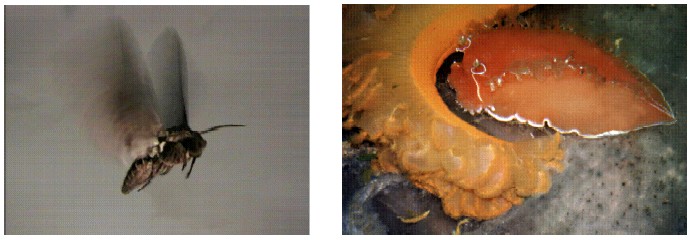
Figure 1: The biological models used in this work. (a) Manduca Sexta is typically 4cm in length, with a 12cm wingspan; at 2.5g, it is among the largest of insect flyers. It can easily carry a test-electronics payload. (b) Tritonia diomedea is typically 20cm in length, and has a readily accessible brain with large and well-characterized neurons. |
|
 |
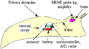 |
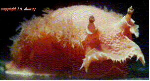
Figure 2: Tritonia is a marine nudibranch foundworldwide with an unusually large species, Tritonia diomedea, indigenous to the Pacific Northwest. Its neuronal cell bodies range from roughly 5µm (the same size as vertebrate neural cells) to specialized cells that are approximately 100 times as large. With our existing experimental platform for this animal we can begin experimenting on the large neurons and then gradually scale to cells of smaller size. (© J. A. Murray) |
Figure 3: Cartoon view of the implantable recording system (not to scale). The microprobe and signal amplifier (total size: 2 x 2 x 1 mm3) are attached to the brain tissue of T. diomedea (the animal is about 20cm in length). A thin tether cable connects to a recording unit in the visceral cavity. The microrecorder comprises a semicustom microcontroller, memory, and battery. |
 |
 |
Figure 4: Evoked extracellular potentials in the lobula plate of Manduca sexta (hawk moth) plotted versus time. |
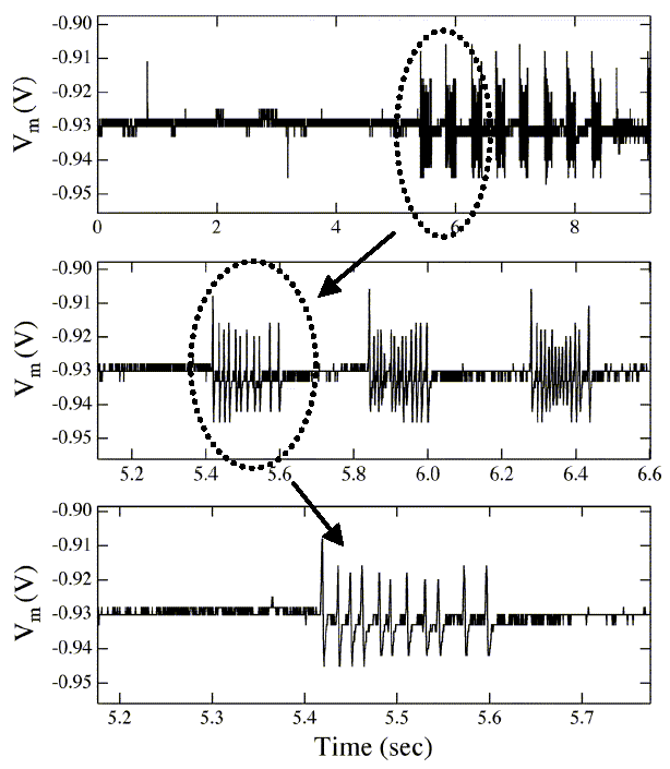
Figure 5: Spontaneous intracellular potentials in a neuron in the brain of Tritonia diomedea (a sea slug) plotted versus time. The positioning of the probe was controlled via micro-manipulators and an optical microscope. |
Selected Publications
-
Yael Hanein, Karl F. Böhringer, Russell C. Wyeth, A. O. Dennis Willows, "Towards MEMS Probes for Intracellular Neuronal Recording," Sensors Update -- Sensor Technology - Applications - Markets 10(1):47-75. Wiley-VHC 2002. Edited by H. Baltes, G. K. Fedder, J. G. Korvink. Invited paper.
A complete list of our publications (many of them available online) can be found here.
Acknowledgements
-
David and Lucile Packard Foundation, grant 2000-01763
-
National Sciences and Research Council (Canada) Post-graduate Scholarship to R. C.W.
-
DARPA Bio:Info:Micro grant MD A972-01-1-002
-
NSF CISE Postdoctoral Research Associateship EIA-0072744 to Y. H.
-
Agilent Technologies, Intel Corporation, Microsoft Research, and Tanner Research Inc.
Sub-micrometer Silicon Needles for Intracellular Applications
Team Members
Yael Hanein, Christian G. J. Schabmueller, Gary Holman, Peter Lücke, Denice D. Denton, Karl F. Böhringer
Summary
A processing technology is presented to produce high-aspect ratio submicrometer silicon needles suited for intracellular interfacing with living cells. Pillars are created using deep reactive ion etching, and the sharpening of the pillars is achieved by reactive ion etching. A simple polyimide-based micro-fabrication approach was developed to integrate the silicon needles with a larger silicon base designed to carry elements such as amplifier, battery or memory. The current design allows convenient handling of the device during implantation and minimal mechanical load on the implanted region. Prototype devices were tested for usability and animal compatibility.
 |
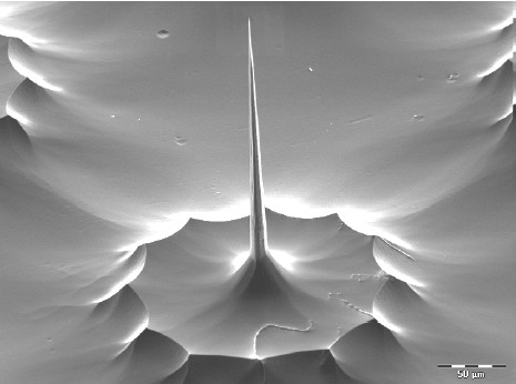 |
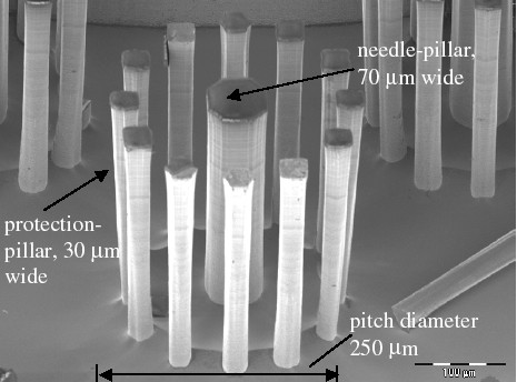
Figure 1: DRIE-etched pillars; pitch diameter 250 µm. The pillars are about 300 µm tall. The use of protection pillars ensures a straight profile. |
Figure 2: RIE-sharpened silicon needle. The needle is 230 µm tall and etched from the needle pillar shown in Figure 1. The protecting pillars are eliminated during the sharpening process. |
Selected Publications
-
Yael Hanein, Christian G. J. Schabmueller, Gary Holman, Peter Luecke, Denice D. Denton, Karl F. Böhringer, "High-Aspect Ratio Submicrometer Needles for Intracellular Applications." IOP Journal of Micromechanics and Microengineering (JMM) 13(4):S91-S95, July 2003. Paper.
A complete list of our publications (many of them available online) can be found here.
Acknowledgements
-
David and Lucile Packard Foundation grant 2000- 01763.
-
DARPA Bio:Info:Micro grant MD A972- 01-1-002
-
NSF CISE Postdoctoral Research Associateship EIA-0072744 to Y.H.
-
Agilent Technologies, Intel Corporation, Microsoft Research and Tanner Research Inc.