
Projects Archive
Biocompatible Coatings for MEMS - Micromachining of Non-fouling Coatings for Bio-MEMS Applications
Team Members
Yael Hanein, Denice Denton, Karl F. Böhringer, Y. Vickie Pan, Buddy D. Ratner (Bioengineering)
Summary
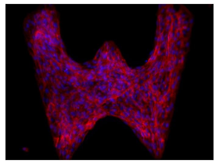
Standard photolithography is used to pattern a poly (ethylene glycol) (PEG)-like polymer onto silicon substrates. The coating has excellent non-fouling properties and good adhesion to various substrate materials, such as silicon, oxide, nitride, gold, and platinum. This method allows precise control of the shape, size, and alignment of the polymer, thus providing a reliable tool to pattern protein sheets as well as cell cultures. This method also enables the incorporation of patterned cell cultures with various predefined elements such as electrodes, channels, and sensors. To demonstrate the properties of our technique, we apply it to build cell cultures and to protect metallic electrodes from pretein and cell adhesion. We show that the thin coatings provide excellent protection without compromising the conductivity of the electrodes.
 |
 |
Figure 1: Micro-patterned cell cultures. Image size approx. 0.5mm. |
Figure 2: Left: electrodes with micromachined and patterned pp4G coating. Right: after incubation in a solution with cells. The pp4G protects the electrodes and prevents cells from sticking. |
Selected Publications
-
Karl F. Böhringer, "Surface Modification and Modulation in Microstructures: Controlling Protein Adsorption, Monolayer Desorption, and Micro-Self-Assembly." IOP Journal of Micromechanics and Microengineering (JMM) 13(4):S1-S10, July 2003. Paper.
-
Yael Hanein, Y. Vickie Pan, Buddy D. Ratner, Denice Denton, Karl F. Böhringer, "Micromachining of Non-fouling Coatings for Bio-MEMS Applications." Sensors and Actuators B: Chemical 81(1):49-54, December 15, 2001. Paper. Posters 1, 2 from UW Undergraduate Research Symposium.
A complete list of our publications (many of them available online) can be found here.
Acknowledgements
-
NSF-ERC EEC-9529161ESCA
-
NIH NCRR grant RR-01296
-
David and Lucile Packard Foundation grant 2000-01763
-
Washington Technology Center awards WTC MEMS 99-8 and 99-A
-
NSF CISE Postdoctoral Research Associateship EIA-0072744 to Y. Hanein
-
NSF Career Award ECS-9875367 to K. Böhringer
-
Microsoft Research, Intel Corporation, and Tanner Research Inc.
Biocompatible Coatings for MEMS - Controlled Microtubules Transport on Patterned Non-adhesive Surfaces
Team Members
Ryan Lipscomb, Yael Hanein, Denice Denton, Karl F. Böhringer, John Clemmens, Viola Vogel, Buddy Ratner (Bioengineering)
Summary
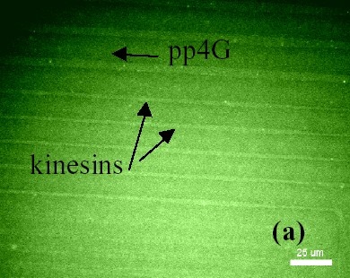
We investigate a lithographic technique used to realize high resolution tracks of the motor protein kinesin inside flow cells. The process consists of patterns of plasma polymerized PEG-like non-fouling coating on a fouling glass substrate. When in contact with a coated surface, proteins adhere exclusively to the fouling areas, thus forming kinesin tracks suited for microtubule guidance. Parallel transport along smooth kinesin track was observed for over 250 µm. A method has been developed to seal the substrates with molded silicon flow channels in order to allow convenient control of reagent introduction and immobilization processes. Complete kinesin and BSA immobilization protocols, comprised of initial rinse, protein introduction, immobilization and final rinse have been successfully demonstrated.
 |
 |
Figure 1: A pp4G pattern on glass with labeled kinesin immobilized on surface along micron-scale tracks. |
Figure 2: Fluorescently labeled microtubules glide on kinesin coated tracks. On regions without kinesin there is no microtubule transport. |
Videos
Selected Publications
-
Ryan C. Lipscomb, John Clemmens, Yael Hanein, Viola Vogel, Buddy D. Ratner, Denice D. Denton and Karl F. Böhringer, "Controlled Microtubules Transport on Patterned Non-adhesive Surfaces." Second International IEEE-EMBS Special Topic Conference on Microtechnologies in Medicine & Biology, pp. 21-26, May 2-4, 2002, Madison, Wisconsin. Paper. Poster.
A complete list of our publications (many of them available online) can be found here.
Acknowledgements
-
David and Lucile Packard Foundation grant 2000-01763
-
NASA grant NAG5- 8784
-
University of Washington Microscale Life Science Center
-
DARPA Bio:Info:Micro grant MD A972-01-1-002
-
NSF CISE Postdoctoral Research Associateship EIA-0072744 to Y.H.
-
Agilent Technologies, Intel Corporation, Microsoft Research and Tanner Research Inc.
-
Center for Nanotechnology IGERT scholarship to J.C.